
And here we are with part two of our rundown on the things you need to learn before you dive into the meaty stuff of A&P, specifically how to talk about the body. In our previous post, we discussed anatomical position and directional terms. In this post, we’re going to take a look at planes and cavities.


No, not the kind that fly you over oceans and have helpful people in uniforms that ply you with bags of stale peanuts. The other kind! The art kind, or in more technical terms the area of a two-dimensional surface. When used in conjunction with anatomy, planes are used to divide the body and its parts, which allows you to describe the views from which you study the body. If you look at your A&P textbook, you’ll most likely notice that a good number of the pictures and diagrams make use of planes.
Here is a list of commonly used planes:
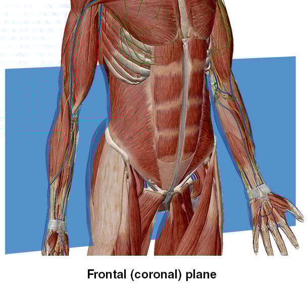
Frontal (Coronal) plane
Divides the body into anterior (front) and posterior (back) portions
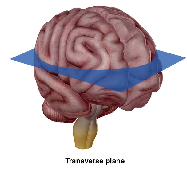
Divides the body into superior (upper) and inferior (lower) portions
Vertical plane that divides the body into right and left sides.
Divides the body at midline into equal right and left sides.
Divides the body at an angle.
Of course, in reality, the planes used are completely imaginary, but they are a helpful visual in terms of describing a view.

Using a frontal plane to bisect the body lengthwise, we’re able to describe certain areas that would not be easily visible or accessible if we used another plane.

The transverse plane bisects the brain horizontally, allowing for a superior view.
Want a quick review of planes, positions, and directional terms? Check out this video:
A concept easier to grasp than planes and directional is body cavities, as they are a physical thing. When you hear the word “cavity,” no doubt you think of the kind in your teeth that are caused by plaque. A cavity, in any capacity, is a hollow place. In your teeth, it’s a hollow bit in the hard body. In the body itself, it is a hollow place usually filled with organs, nerves, vessels, and muscles.
Here are the body’s cavities:
Formed by the cranial bones and holds the brain
Formed by the vertebrae and contains the spinal cord
Formed by the thoracic cage, muscles of the chest, sternum, and the thoracic vertebrae; contains the pleural, pericardial, and mediastinum cavities
Fluid-filled spaces that surround both lungs
Fluid-filled space that surrounds the heart; the serous membrane of the pericardial cavity is the pericardium
Central portion of the thoracic cavity; contains the heart, thymus, trachea, several major blood vessels, and esophagus
Contains liver, stomach, spleen, small intestine, and most of the large intestine; the serous membrane of the abdominal cavity is the peritoneum
Contains bladder, some of the large intestine, and reproductive organs (internal)

The cranial cavity. Image from Visible Body Suite.

The thoracic cavity. Image from Visible Body Suite.

The abdominal cavity. Image from Visible Body Suite.

The pelvic cavity. Image from Visible Body Suite.
This post was originally published in 2013. It has since been updated with new body cavity images from VB Suite.

Be sure to subscribe to the Visible Body Blog for more anatomy awesomeness!
Are you an instructor? We have award-winning 3D products and resources for your anatomy and physiology course! Learn more here.